In An Ekg The P Wave Is Generated When The
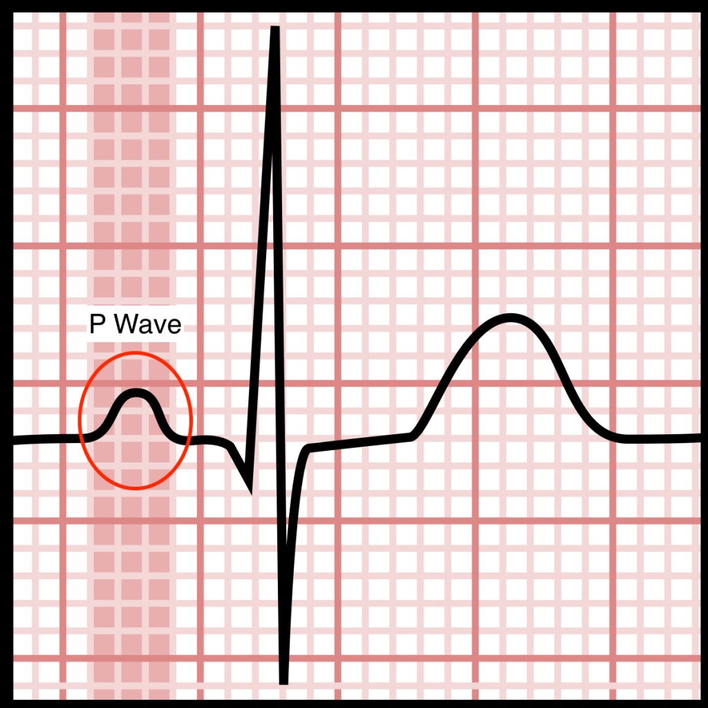
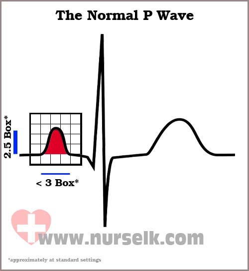
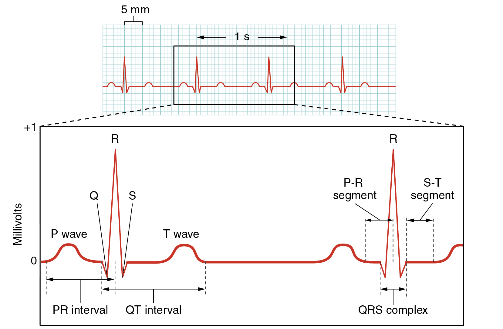
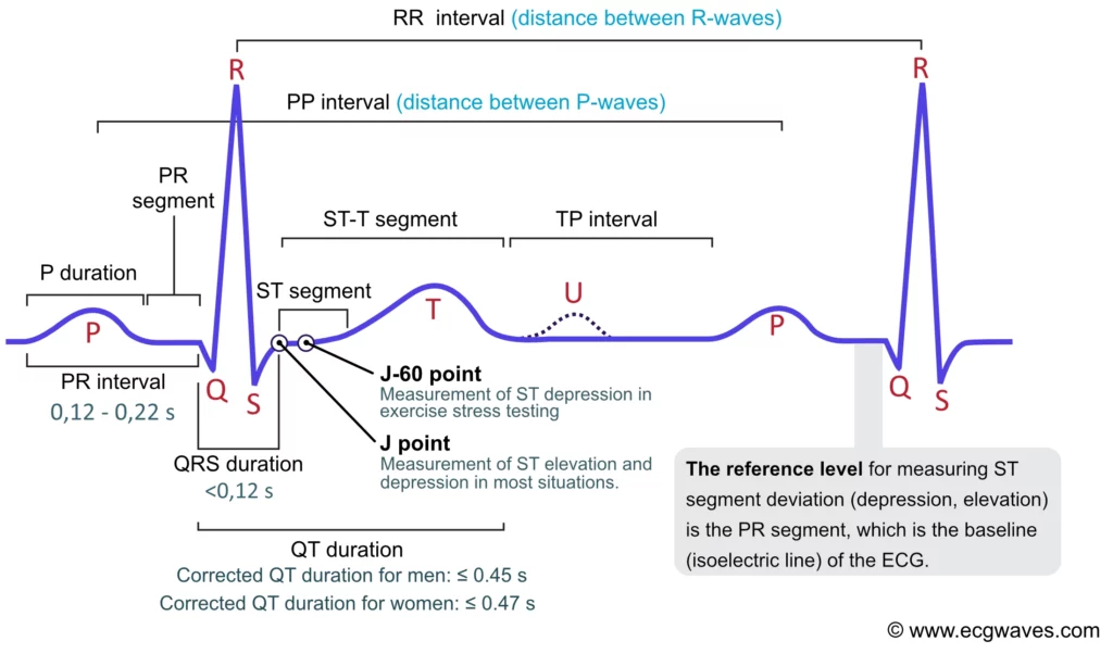
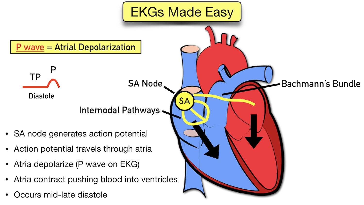
In An Ekg The P Wave Is Generated When The - The presence of multiple p wave. Match the component of the electrocardiogram to the correct definition. When the pr interval is ≥ 120 ms, the origin is within the atria (e.g. The right atrium (ra) is depolarized towards the av node. The p wave represents atrial depolarization, which starts in the sinus node.
The right atrium (ra) is depolarized towards the av node. The p wave represents atrial depolarization, which starts in the sinus node. When the pr interval is ≥ 120 ms, the origin is within the atria (e.g. Match the component of the electrocardiogram to the correct definition. The presence of multiple p wave.
When the pr interval is ≥ 120 ms, the origin is within the atria (e.g. The p wave represents atrial depolarization, which starts in the sinus node. The right atrium (ra) is depolarized towards the av node. Match the component of the electrocardiogram to the correct definition. The presence of multiple p wave.
Electrocardiograms (EKGs/ECGs) Evaluating P Waves Stepwards
The presence of multiple p wave. When the pr interval is ≥ 120 ms, the origin is within the atria (e.g. The right atrium (ra) is depolarized towards the av node. The p wave represents atrial depolarization, which starts in the sinus node. Match the component of the electrocardiogram to the correct definition.

Medical School Qrs complex, Ekg, P wave
The p wave represents atrial depolarization, which starts in the sinus node. The presence of multiple p wave. The right atrium (ra) is depolarized towards the av node. Match the component of the electrocardiogram to the correct definition. When the pr interval is ≥ 120 ms, the origin is within the atria (e.g.

P wave, QRS complex and T wave. Cardiac physiology Nursing study
The p wave represents atrial depolarization, which starts in the sinus node. Match the component of the electrocardiogram to the correct definition. The presence of multiple p wave. When the pr interval is ≥ 120 ms, the origin is within the atria (e.g. The right atrium (ra) is depolarized towards the av node.

The Cardiac Cycle on an EKG (PQRST Waves) YouTube
The p wave represents atrial depolarization, which starts in the sinus node. Match the component of the electrocardiogram to the correct definition. The right atrium (ra) is depolarized towards the av node. When the pr interval is ≥ 120 ms, the origin is within the atria (e.g. The presence of multiple p wave.
How to read an Electrocardiogram (ECG) Part 3, The P Wave
The p wave represents atrial depolarization, which starts in the sinus node. Match the component of the electrocardiogram to the correct definition. When the pr interval is ≥ 120 ms, the origin is within the atria (e.g. The presence of multiple p wave. The right atrium (ra) is depolarized towards the av node.
19.2 Cardiac Muscle and Electrical Activity Douglas College Human
The right atrium (ra) is depolarized towards the av node. Match the component of the electrocardiogram to the correct definition. The presence of multiple p wave. When the pr interval is ≥ 120 ms, the origin is within the atria (e.g. The p wave represents atrial depolarization, which starts in the sinus node.

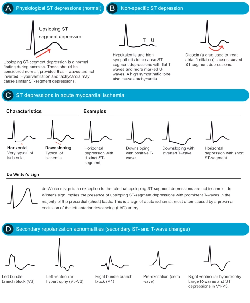
ECG interpretation Characteristics of the normal ECG (Pwave, QRS
The presence of multiple p wave. Match the component of the electrocardiogram to the correct definition. The p wave represents atrial depolarization, which starts in the sinus node. The right atrium (ra) is depolarized towards the av node. When the pr interval is ≥ 120 ms, the origin is within the atria (e.g.
ECG Waveform Explained EKG Labeled Diagrams And Components, 60 OFF
The p wave represents atrial depolarization, which starts in the sinus node. Match the component of the electrocardiogram to the correct definition. The presence of multiple p wave. When the pr interval is ≥ 120 ms, the origin is within the atria (e.g. The right atrium (ra) is depolarized towards the av node.
ECG Interpretation Characteristics Of The Normal ECG, 57 OFF
The presence of multiple p wave. Match the component of the electrocardiogram to the correct definition. The right atrium (ra) is depolarized towards the av node. When the pr interval is ≥ 120 ms, the origin is within the atria (e.g. The p wave represents atrial depolarization, which starts in the sinus node.
ECG Waveform Explained EKG Labeled Diagrams And Components
When the pr interval is ≥ 120 ms, the origin is within the atria (e.g. The right atrium (ra) is depolarized towards the av node. The presence of multiple p wave. Match the component of the electrocardiogram to the correct definition. The p wave represents atrial depolarization, which starts in the sinus node.
The Right Atrium (Ra) Is Depolarized Towards The Av Node.
The presence of multiple p wave. Match the component of the electrocardiogram to the correct definition. The p wave represents atrial depolarization, which starts in the sinus node. When the pr interval is ≥ 120 ms, the origin is within the atria (e.g.