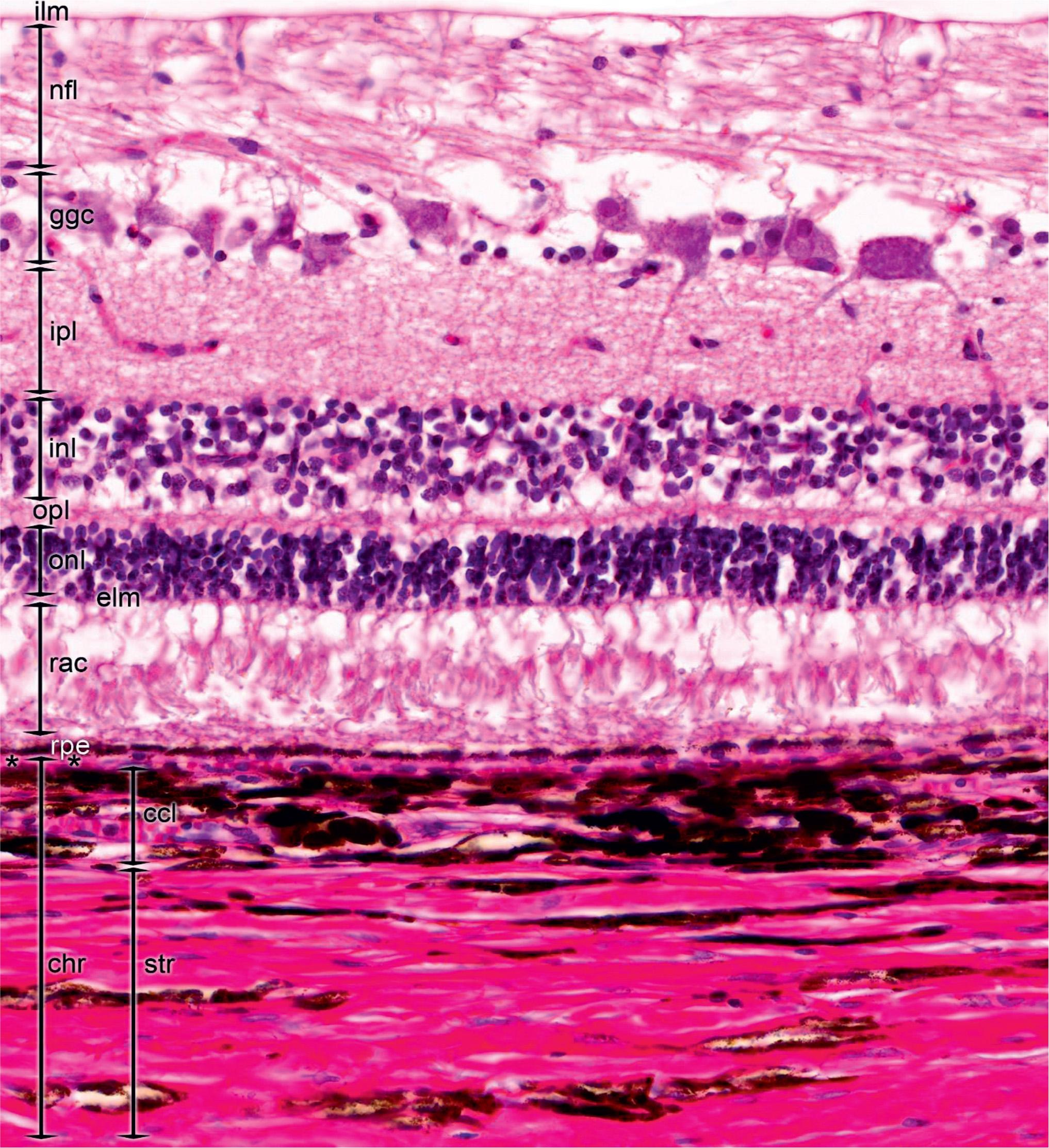
Forms The Bulk Of The Heavily Pigmented Vascular Layer
Forms The Bulk Of The Heavily Pigmented Vascular Layer - In the neural layer, the neuron populations are arranged as follows. Forms the bulk of the heavily pigmented vascular layer: The two major layers of the retina are the pigmented and neural layers. Study with quizlet and memorize flashcards containing terms like fluid. Composed of tough, white, opaque, fibrous connective tissue. This layer provides a blood supply to the eyeball. This layer is richly supplied with blood vessels and. It delivers oxygen and nutrients to the outer layers. The choroid plexuses form the bulk of the heavily pigmented vascular layer in the eye. The bulk of the heavily pigmented vascular layer of the eye is formed by the choroid.
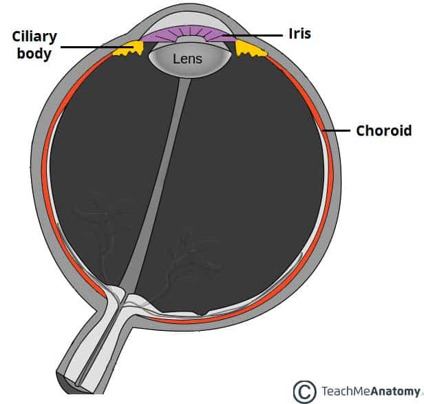
Forms the bulk of the heavily pigmented vascular layer: In the neural layer, the neuron populations are arranged as follows. This layer is richly supplied with blood vessels and. Study with quizlet and memorize flashcards containing terms like aqueous humor, sclera, optic disc and more. This layer provides a blood supply to the eyeball. The two major layers of the retina are the pigmented and neural layers. The bulk of the heavily pigmented vascular layer of the eye is formed by the choroid. The choroid, a heavily pigmented vascular layer, forms the largest part of the eye. The choroid plexuses form the bulk of the heavily pigmented vascular layer in the eye. It delivers oxygen and nutrients to the outer layers.
Study with quizlet and memorize flashcards containing terms like aqueous humor, sclera, optic disc and more. Forms the bulk of the heavily pigmented vascular layer: In the neural layer, the neuron populations are arranged as follows. It delivers oxygen and nutrients to the outer layers. The choroid, a heavily pigmented vascular layer, forms the largest part of the eye. This layer provides a blood supply to the eyeball. Composed of tough, white, opaque, fibrous connective tissue. The two major layers of the retina are the pigmented and neural layers. The bulk of the heavily pigmented vascular layer of the eye is formed by the choroid. The choroid plexuses form the bulk of the heavily pigmented vascular layer in the eye.
Sensory Organs Clinical Tree
Study with quizlet and memorize flashcards containing terms like fluid. This layer is richly supplied with blood vessels and. Forms the bulk of the heavily pigmented vascular layer: The choroid plexuses form the bulk of the heavily pigmented vascular layer in the eye. In the neural layer, the neuron populations are arranged as follows.

There are three layers, or tunics, of the eyeball. The fibrous layer is
In the neural layer, the neuron populations are arranged as follows. Study with quizlet and memorize flashcards containing terms like aqueous humor, sclera, optic disc and more. Composed of tough, white, opaque, fibrous connective tissue. The choroid, a heavily pigmented vascular layer, forms the largest part of the eye. Forms the bulk of the heavily pigmented vascular layer:
Vascular tunic neurolader
Study with quizlet and memorize flashcards containing terms like fluid. It delivers oxygen and nutrients to the outer layers. Composed of tough, white, opaque, fibrous connective tissue. The bulk of the heavily pigmented vascular layer of the eye is formed by the choroid. The choroid plexuses form the bulk of the heavily pigmented vascular layer in the eye.
+Tunic.jpg)
Vision Interactive pgs ppt download
This layer is richly supplied with blood vessels and. Study with quizlet and memorize flashcards containing terms like aqueous humor, sclera, optic disc and more. The choroid, a heavily pigmented vascular layer, forms the largest part of the eye. It delivers oxygen and nutrients to the outer layers. The two major layers of the retina are the pigmented and neural.

Pigmentation Biology for Majors II
Study with quizlet and memorize flashcards containing terms like fluid. Study with quizlet and memorize flashcards containing terms like aqueous humor, sclera, optic disc and more. The bulk of the heavily pigmented vascular layer of the eye is formed by the choroid. This layer provides a blood supply to the eyeball. The two major layers of the retina are the.

Retina 4 Digital Histology
Study with quizlet and memorize flashcards containing terms like fluid. The two major layers of the retina are the pigmented and neural layers. Study with quizlet and memorize flashcards containing terms like aqueous humor, sclera, optic disc and more. In the neural layer, the neuron populations are arranged as follows. The choroid, a heavily pigmented vascular layer, forms the largest.

Xylem Wikipedia in 2020 Tissue types, Tissue biology, Biology notes
The bulk of the heavily pigmented vascular layer of the eye is formed by the choroid. The choroid, a heavily pigmented vascular layer, forms the largest part of the eye. Forms the bulk of the heavily pigmented vascular layer: In the neural layer, the neuron populations are arranged as follows. This layer is richly supplied with blood vessels and.
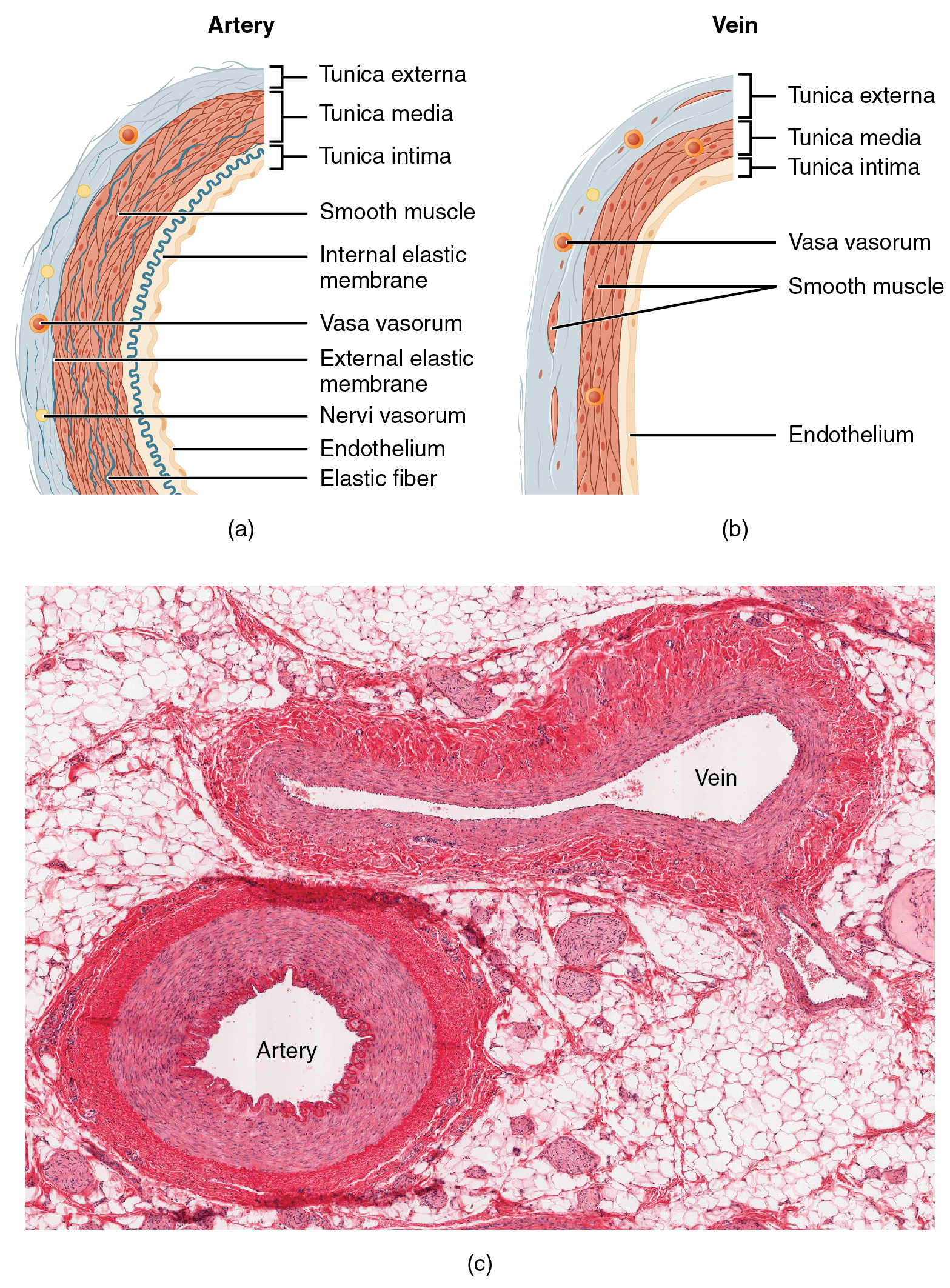
20.1 Structure and Function of Blood Vessels Douglas College Human
In the neural layer, the neuron populations are arranged as follows. Study with quizlet and memorize flashcards containing terms like fluid. This layer provides a blood supply to the eyeball. It delivers oxygen and nutrients to the outer layers. The choroid plexuses form the bulk of the heavily pigmented vascular layer in the eye.

Sensory systems online presentation
The two major layers of the retina are the pigmented and neural layers. The choroid plexuses form the bulk of the heavily pigmented vascular layer in the eye. The choroid, a heavily pigmented vascular layer, forms the largest part of the eye. In the neural layer, the neuron populations are arranged as follows. Forms the bulk of the heavily pigmented.

Vascular Pigmented Layer PDF Human Eye Cornea
The choroid plexuses form the bulk of the heavily pigmented vascular layer in the eye. Study with quizlet and memorize flashcards containing terms like fluid. This layer provides a blood supply to the eyeball. Composed of tough, white, opaque, fibrous connective tissue. Study with quizlet and memorize flashcards containing terms like aqueous humor, sclera, optic disc and more.
Study With Quizlet And Memorize Flashcards Containing Terms Like Fluid.
Forms the bulk of the heavily pigmented vascular layer: The two major layers of the retina are the pigmented and neural layers. Composed of tough, white, opaque, fibrous connective tissue. It delivers oxygen and nutrients to the outer layers.
The Choroid Plexuses Form The Bulk Of The Heavily Pigmented Vascular Layer In The Eye.
The choroid, a heavily pigmented vascular layer, forms the largest part of the eye. The bulk of the heavily pigmented vascular layer of the eye is formed by the choroid. Study with quizlet and memorize flashcards containing terms like aqueous humor, sclera, optic disc and more. This layer provides a blood supply to the eyeball.
This Layer Is Richly Supplied With Blood Vessels And.
In the neural layer, the neuron populations are arranged as follows.